A pathology slide — a sliver of tumor tissue on a glass slide, stained and viewed under a microscope — has always been read for one basic question: is this cancer, and if so, what kind. At the American Association for Cancer Research’s 2026 annual meeting in San Diego, where more than 7,000 abstracts were presented, several research groups showed something more ambitious: AI models that look at the same ordinary stained slides and predict genetic mutations and treatment responses that doctors have historically needed separate, expensive molecular tests to find.
The Colorectal Cancer Model That Hit 98% Accuracy
Genetic testing company Natera presented a deep learning model trained on paired molecular and histopathology data from more than 45,000 colorectal cancer patients. Critically, once trained, the model needs only the standard H&E-stained slide — no additional sequencing — to make its predictions at the point of use. In testing, it predicted microsatellite instability (MSI) status, a biomarker that determines whether a colorectal tumor is likely to respond to immunotherapy, with 0.98 accuracy, and predicted BRAF V600E mutation status, which affects treatment selection, with 0.93 accuracy.
MD Anderson’s Model Says It Beats the Standard Biomarker Test
A second project, Path-IO, developed by researchers at MD Anderson Cancer Center, was validated on more than 1,000 patients across multiple institutions and countries. It predicts how a non-small cell lung cancer patient will respond to immunotherapy using whole-slide images and pathology data, and the team reported it outperformed PD-L1 testing — the standard biomarker test oncologists currently use to help decide who gets immunotherapy — across its validation datasets. The presenters didn’t publish the specific head-to-head numbers in the materials reviewed, which is worth flagging before treating “outperforms PD-L1” as settled.
Reading Gene Activity Off a Stained Slide
A third effort, called Path2Omics or TIME_ACT, came from a multi-institution team spanning the National Cancer Institute, Cedars-Sinai, Harvard Medical School, Dana-Farber Cancer Institute, and Yale. Their model infers gene expression directly from H&E images, identifying activity across 66 genes linked to tumor immune activation without sequencing, extra staining, or any molecular testing at all — essentially trying to read a genomic signal out of a picture that was never designed to carry one.
Why Any of This Matters Beyond the Conference Room
The case for this approach rests on a few practical realities. Tissue biopsies are often small, and every slice used for a molecular test is tissue that can’t be used for something else — AI analysis consumes none of it. Molecular testing is also expensive and slow, which matters most in exactly the settings — under-resourced hospitals, lower-income countries — where patients can least afford the wait. And because every oncology trial participant already has at least one associated pathology slide on file, researchers argue there’s a vast, underused archive of digitized and digitizable slides that could be mined retrospectively for biomarkers researchers didn’t even know to look for when the slides were first taken. As the pathology AI company Proscia put it in a write-up of the conference, “pathology data offers the most detailed and direct profile of disease.”
The Skeptic’s Checklist
Nearly all of this is conference-stage material, not yet published, peer-reviewed findings sitting in a journal where independent experts have picked apart the methodology. Natera’s 0.98 and 0.93 accuracy figures, MD Anderson’s claim of beating PD-L1, and the 66-gene signature from Path2Omics all need to survive that scrutiny, plus prospective validation in new patient populations the models weren’t trained or tuned on. There’s also a basic infrastructure problem: before any of these models can be used broadly, archived slides have to be digitized, annotated, and curated, which is slow and expensive work that institutions have historically underinvested in. And even a model that reliably predicts a mutation from a slide still needs a regulatory pathway — likely as a companion diagnostic, the same category that governs tests used to decide which drug a patient gets — before it can replace, rather than merely supplement, the lab tests oncologists currently order.
The Road From Conference Poster to Clinic
If these models hold up under independent, peer-reviewed validation, the more interesting downstream effect may not be at the bedside but in drug development: pharmaceutical companies running oncology trials could use AI-derived biomarkers to identify and enroll the patients most likely to respond to an experimental drug, without waiting on sequencing turnaround times. For a tool to go from predicting a mutation to actually changing what drug a specific patient receives, though, it will need the kind of large, prospective, multi-institution trial that none of the AACR 2026 presentations described yet — the gap between a striking conference accuracy number and an FDA-cleared companion diagnostic remains wide.
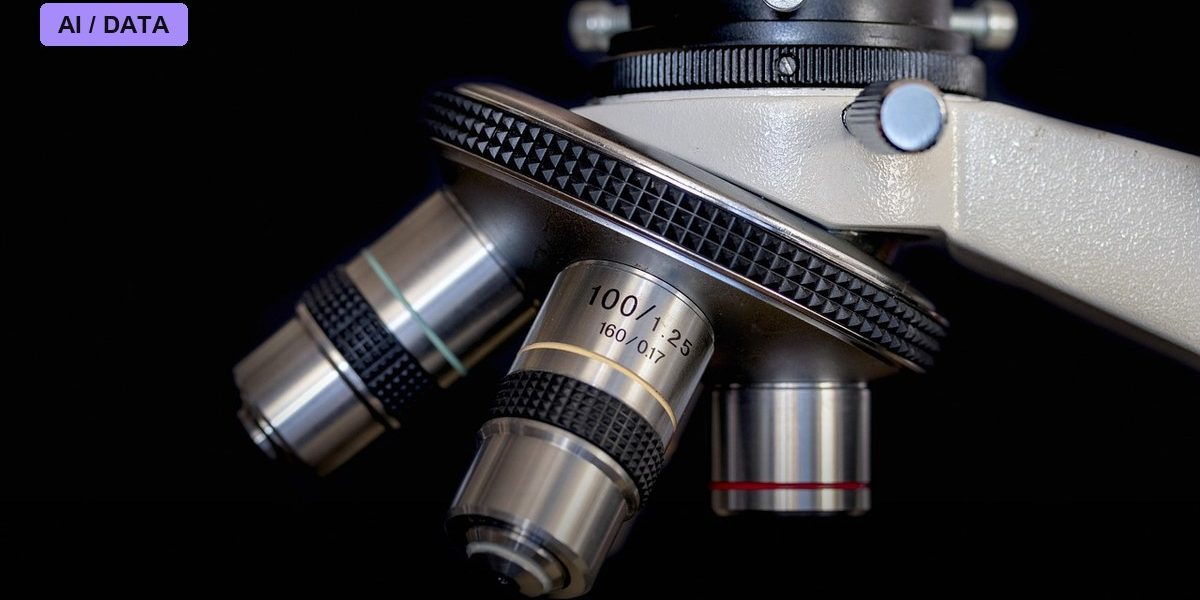
Photo: Panals / PIXABAY via Pixabay