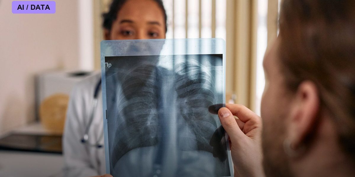
Osteoporosis is famous for being a silent disease until it isn’t — the first clear sign is often a fracture. A study presented at the World Congress on Osteoporosis in April 2026 argues that one of the most common medical images already being taken, the routine chest X-ray, can be repurposed by AI to catch dangerously low bone density years before anyone orders a dedicated bone scan.
A Scan Everyone Already Gets
The gold standard for diagnosing osteoporosis is a DXA scan, a dedicated low-radiation X-ray that measures bone mineral density directly. The problem, researchers note, is that DXA access and uptake vary enormously depending on where a patient lives and whether a doctor thinks to order one in the first place, which means osteoporosis often goes undiagnosed until a hip or spine fracture forces the issue — at which point the clinical and financial cost is far higher. Chest X-rays, by contrast, are taken constantly, for pneumonia checks, pre-surgical clearance, routine physicals, and dozens of other reasons that have nothing to do with bone health. The research team behind a tool called OsteoPredict set out to see whether those already-captured images contain enough signal to flag low bone density on their own.
How OsteoPredict Was Built and Tested
In an abstract titled “Multi-Center Validation of a Federated Learning-Based Artificial Intelligence System for Screening Low Bone Mineral Density Using Chest X-Rays,” presented by lead author Hung WC and colleagues, the team trained the model on nearly 6,000 chest X-ray images using federated learning — a technique that lets a model learn from data held at multiple institutions without that data ever leaving each site. They then externally validated it on more than 3,000 patients drawn from two independent medical centers that hadn’t contributed training data. The tool is designed to flag patients likely to have a T-score of -2.5 or lower, the clinical threshold that defines osteoporosis.
The Performance Numbers
OsteoPredict posted an area under the curve (AUROC) of 0.942, with 91.0% sensitivity and 82.7% specificity in external validation — strong enough that the researchers reported the performance held up consistently across both validation sites and between male and female patients. Notably, sensitivity remained high in patients 60 and older, the age group where osteoporosis risk and fracture consequences are both highest. A separate but related effort, from researchers at St. Paul’s Hospital and National Taiwan University, repurposed routine chest X-rays taken during general health exams and found something striking: more than half of the confirmed abnormal bone-density cases they identified occurred in patients with a normal body mass index — exactly the kind of patient that standard risk-based screening questionnaires are prone to miss, since low BMI is a known osteoporosis risk factor that higher-BMI patients don’t trigger.
A Second Opinion From Hip X-Rays
OsteoPredict isn’t the only opportunistic bone-density tool moving through the pipeline. OsteoSight, developed by Naitive Technologies, works on hip and pelvis X-rays instead of chest images, received FDA clearance in 2025, and integrates directly into electronic health records so flags show up where clinicians are already working. Research on it was presented at the 2026 American Academy of Orthopaedic Surgeons annual meeting, and a 2025 study in Osteoporosis International validated it against a US patient population. The parallel development of multiple opportunistic-screening tools, built on different scan types, suggests the field sees real appetite for catching osteoporosis through images clinicians already have on file rather than ordering new tests.
Reasons for Caution
The researchers behind OsteoPredict were upfront about the limits of their own study: it was retrospective, built and tested on images already collected, not on a forward-looking clinical workflow where a flag actually changes what a doctor does next. Prospective validation — tracking what happens when real clinicians receive real-time AI flags on real chest X-rays — hasn’t happened yet. There are also open questions about what a positive flag should trigger: a confirmatory DXA scan is an additional cost and appointment, and health systems would need to work out who pays for that follow-up and whether catching more at-risk patients earlier actually proves cost-effective once you account for the false positives an 82.7% specificity rate implies.
What Comes Next
The appeal of opportunistic screening is that it doesn’t ask patients or health systems to do anything new — it just asks existing images to work harder. If prospective trials confirm that AI flags on routine chest X-rays reliably catch osteoporosis that would otherwise go undetected until a fracture, the more interesting fight will be over implementation: whether radiology departments bolt this kind of screening onto every chest X-ray by default, whether insurers will cover the DXA follow-up it generates, and whether primary care physicians trust an AI flag enough to act on it. For now, the WCO 2026 data is a proof of concept, not yet a standard of care.
Photo: cottonbro studio / PEXELS via Pexels