Sutter Health, the Northern California nonprofit hospital network that performs roughly 370,000 screening mammograms a year, says an artificial intelligence platform it rolled out across its imaging centers is finding more cancers without generating more false alarms. In the second quarter of 2025 alone, the health system ran AI analysis on more than 35,000 screening mammograms, and administrators say the breast cancer detection rate climbed from a baseline of 4.8 cases per 1,000 screenings to more than 6.0 per 1,000 — a jump large enough that Sutter has now folded the technology into its system-wide Breast Cancer Programs of Oncology Distinction.
How The Technology Works
The AI layer running underneath Sutter’s mammography workflow comes from Ferrum Health, a health care AI platform company whose software flags areas of concern on a scan before or alongside a radiologist’s own read, rather than replacing that read. Sutter’s figures show the tool is doing more than spotting tumors: it also flagged 14% of women screened as high-risk for future breast cancer and 7% as extremely high-risk, categories that can trigger earlier or more frequent follow-up imaging. Separately, the software identified dense breast tissue in 38% of patients — a known risk factor that makes tumors harder to see on a standard mammogram — while 44% of studies came back with no AI-flagged areas at all, underscoring that the tool is being used as a triage and second-check layer rather than a blanket cancer-detection trigger.
Why Detection Accuracy Is Such a Stubborn Problem
Mammography has always had a known blind spot: radiologists reading scans under time pressure can miss small or subtle tumors, particularly in dense breast tissue, and those misses sometimes surface later as fast-growing interval cancers that develop between scheduled screenings. Academic studies of AI-assisted mammography have put rough numbers on that gap — one widely cited UCLA-led analysis estimated AI could help cut the number of interval cancers missed at initial screening by roughly 30%, and a Swedish trial found AI-supported reading produced a cancer detection rate about 20% higher than human-only reading, without increasing the false-positive rate. Sutter’s internal numbers, while not a peer-reviewed clinical trial, land in a similar range and reflect a pattern showing up across health systems that have adopted AI mammography support over the past two years.
The People Behind the Push
“By expanding access and investing in innovation, we’re redefining cancer care from prevention to survivorship, today and for the future,” said Dr. Nitin Rohatgi, a medical oncologist who chairs Sutter Health’s Breast Cancer Programs of Oncology Distinction. Ferrum Health co-founder and chief executive Pelu Tran has pitched the broader pattern similarly to health systems nationally, positioning AI not as an autonomous diagnostician but as a tool that reviews every scan the same way, every time, catching findings a tired or rushed reader might skip. Sutter has pushed the same software into a mobile mammography van, which has screened nearly 500 women since its spring 2025 launch and flagged 12% as high-risk and 3% as extremely high-risk — numbers Sutter cites as evidence the tool performs consistently even in a lower-resource, community-outreach setting rather than only in a fully staffed hospital radiology suite.
A Trend, Not an Isolated Case
Sutter isn’t an outlier. Mount Sinai Health System in New York disclosed in March 2025 that it had performed more than 100,000 AI-assisted mammograms, with Dr. Laurie Margolies, chief of breast imaging at the system’s Dubin Breast Center, telling the health system’s newsroom that “artificial intelligence is a phenomenal tool” that “does not replace the expertise of our radiologists — it enhances it.” Regulators have been approving the underlying software at a steady clip: the FDA cleared DeepHealth’s Saige-Density tool, which automatically classifies breast density using the American College of Radiology’s BI-RADS scale, after a multicenter study found 91.5% agreement with human specialist assessments, and separately cleared an updated version of Lunit’s 3D mammography algorithm that can compare a new scan against up to two prior exams. RadNet, the country’s largest outpatient imaging chain, moved to acquire AI mammography developer iCAD in a roughly $103 million deal to make the technology standard across its centers.
The Skeptics’ Case
Not every study paints an unqualified win. South Korea’s AI-STREAM trial, a large multicenter comparison, found that AI computer-aided detection software caught about 3.4% of cancers that radiologists initially missed — but the same software missed 8.1% of cancers that radiologists caught on their own recall, with overall sensitivity landing at 89.9% and specificity at 94.3%. A separate population-based screening study comparing AI and radiologist false positives on tomosynthesis exams found nearly identical overall false-positive rates of about 10% for both, but with strikingly different error patterns: 43% of false positives came from AI alone and 44% from radiologists alone, meaning AI is not simply a strictly-better version of a human reader but a tool that catches different things and misses different things. Critics also point to equity concerns — AI models trained predominantly on data from one population or imaging equipment vendor may perform less reliably on other demographic groups or on different scanner hardware, a gap regulators and hospital systems are still working to measure systematically.
What Comes Next
The near-term trajectory looks like deeper integration rather than a single breakthrough moment: more health systems pairing AI density and risk-scoring tools with routine screening, more FDA clearances for narrower applications like calcification detection and prior-exam comparison, and ongoing trials — including an active U.S. study comparing single-radiologist-plus-AI reading against traditional double reading by two radiologists — aimed at establishing whether AI can safely reduce the workforce burden on a radiology specialty that is already short-staffed in many regions. For patients, the practical change is already visible in facilities like Sutter’s: a scan that gets read once by a radiologist and, invisibly, a second time by software trained to catch exactly the kind of subtle finding that produces a late-stage diagnosis down the road.
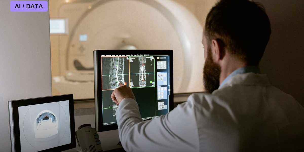
Photo: MART PRODUCTION / PEXELS via Pexels