Researchers at MIT’s Computer Science and Artificial Intelligence Laboratory, working with collaborators at Harvard Medical School and Massachusetts General Hospital, have built an AI system that could make minimally invasive surgery safer and more widely available by solving a problem that has quietly limited how many hospitals can perform the most precise image-guided procedures. The work, published in Nature on September 16, 2026, describes a technique called xvr — short for X-ray volume registration — that takes a single 2D X-ray captured mid-surgery and matches it to a patient’s preoperative 3D CT or MRI scan in a matter of seconds, with sub-millimeter accuracy.
The problem xvr is trying to solve
During many modern surgical procedures — particularly minimally invasive orthopedic and neurosurgical operations — surgeons rely on live X-ray imaging to see where their instruments are inside the body in real time. But a single 2D X-ray is a flattened projection; it doesn’t tell a surgeon exactly how that view lines up with the detailed 3D map of the patient’s anatomy captured before surgery. Translating between the two views, known as image registration, has traditionally required either specialized hardware, time-consuming manual alignment, or algorithms that struggle to generalize across different patients, body parts and procedures. That bottleneck has limited how broadly the most image-intensive minimally invasive techniques can be deployed, particularly in hospitals without the most advanced imaging suites.
How the AI actually works
The MIT-led team’s approach is built around a clever training trick: rather than relying on scarce real-world paired X-ray and 3D scan data, xvr generates thousands of synthetic X-rays from a patient’s existing 3D scan using physics-based simulation of how X-rays pass through tissue and bone. It then trains a model on those synthetic images to recognize how a real, live X-ray taken during surgery should align with the original 3D scan. According to the research team, led by CSAIL postdoctoral researcher Vivek Gopalakrishnan and MIT professor Polina Golland, along with Harvard Medical School and Mass General investigator Neel Dey, the resulting system can register a real surgical X-ray to a patient’s 3D anatomy in about five minutes of training time and then perform the alignment itself in seconds, fast enough to be usable even in emergency surgical settings.
How big of an improvement this represents
The team reports that xvr outperformed existing AI-based registration methods “by an order of magnitude” in accuracy and robustness, tested across multiple patients, body parts and procedure types — a notably broad validation compared to prior tools that often work well only for the narrow anatomical region or imaging setup they were built and tuned for. That generalizability is central to the researchers’ pitch: a registration tool that only works reliably for spine surgery, for instance, does little to help a neurosurgeon performing a completely different procedure. The Nature paper’s co-authors also included researchers and clinicians from Boston Children’s Hospital and Shriners Children’s Hospital, hospitals whose pediatric surgical teams often work with more complex anatomical variation than adult-focused surgical centers.
Why this matters beyond the operating room where it was tested
Gopalakrishnan framed the significance of the work in terms of access rather than just precision, saying that “making these procedures easier by combining 2D and 3D information enables these types of highly specialized life-saving procedures to be more accessible to much broader populations.” That framing points to a persistent inequity in surgical care: the most image-guided, minimally invasive techniques — which generally mean less trauma, shorter recovery times and fewer complications for patients — have historically been concentrated in major academic medical centers with the imaging infrastructure and specialized staff to support them. A registration tool that runs fast and generalizes well, without requiring exotic hardware, could in principle let smaller hospitals offer some of the same image-guided precision that has been the province of large surgical centers.
What still has to happen before this reaches patients
Publication in Nature establishes the scientific credibility of the underlying method, but xvr is a research result, not yet a commercially deployed, FDA-cleared surgical tool. Moving from an academic paper with strong benchmark results to something integrated into an operating room’s imaging systems typically requires additional validation studies, regulatory clearance, and partnerships with medical device manufacturers who build the C-arm X-ray machines and surgical navigation platforms hospitals actually use. The research team has not announced a commercialization timeline or an industry partner to bring xvr to market. Researchers in the broader surgical AI field have also cautioned that techniques validated at leading academic hospitals like Mass General and Boston Children’s don’t always transfer cleanly to the more variable equipment and patient populations found in community hospitals, which is precisely the audience the technology is meant to serve.
What’s next
The MIT team’s next steps are likely to involve prospective clinical validation — testing xvr in live surgical settings rather than retrospective analysis of existing imaging data — and conversations with device makers about integrating the registration engine into existing surgical navigation hardware. Given the broad interest from Boston-area hospitals already involved in the research, expect follow-up studies focused on specific surgical specialties, such as spine or orthopedic trauma procedures, where fast, accurate 2D-to-3D registration would have the most immediate clinical payoff.
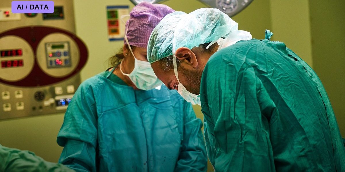
Photo: Engin_Akyurt / PIXABAY via Pixabay