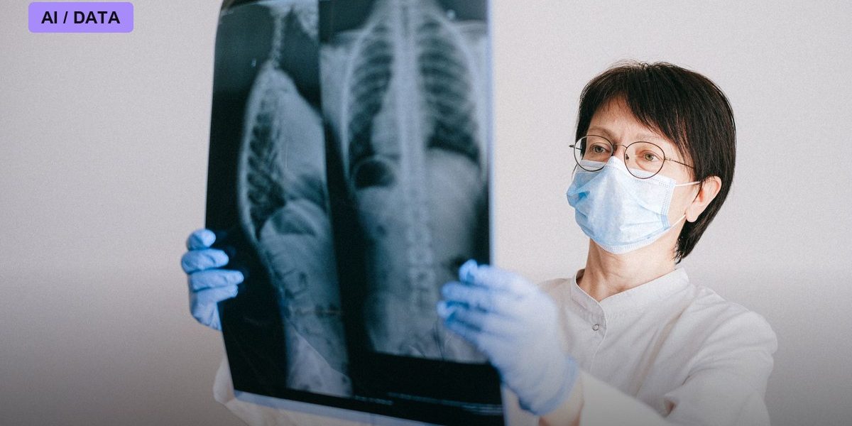
A chest X-ray is one of the most ordinary tests in medicine — ordered for coughs, pre-surgery checklists, and pneumonia scares, then usually filed away and forgotten. A study published April 1, 2026, in the journal Radiology: Artificial Intelligence suggests those routine images are quietly encoding something doctors have never been able to read directly: how fast a person’s body is actually aging, independent of the number on their birth certificate.
A model trained to guess your age from your bones and lungs
Researchers led by Yoosoo Chang, MD, PhD, of Sungkyunkwan University and Hyungjin Kim, MD, PhD, of Seoul National University built a deep learning model called AgeNet and trained it on 111,266 chest X-rays from 64,254 asymptomatic adults between the ages of 40 and 80. The model’s job was narrow: look at a chest X-ray and guess the patient’s chronological age based purely on radiographic features — bone density, cardiac silhouette, lung architecture, soft tissue patterns. Once trained, the researchers applied AgeNet to a much larger population: 421,894 healthy Korean adults whose chest X-rays were collected between January 2006 and December 2020, then tracked for a median of 8.5 years to see who died and from what.
When the X-ray says you’re older than you are
The core finding is that the gap between a person’s AI-estimated “radiographic age” and their actual chronological age predicts mortality. Patients whose AI-estimated age exceeded their real age — termed “accelerated aging” — had meaningfully higher death rates during follow-up: a hazard ratio of 1.26 for men and 1.52 for women, meaning accelerated-aging women in the study had roughly 52 percent higher mortality risk than their peers with normal or decelerated aging scores. Over the study period, 6,506 participants died, split across 953 cardiovascular deaths, 3,024 cancer deaths, and 1,043 respiratory deaths. A follow-up analysis that tracked people with three or more X-rays over time — covering 179,667 participants — went further, measuring not just a single age gap but the velocity of aging. People whose radiographic age accelerated fastest over repeated scans had mortality rate ratios of 1.51 for men and 1.71 for women, while those whose radiographic aging actually slowed down had sharply lower mortality risk, as low as 0.50 for women relative to average agers.
What the AI is actually picking up on
The researchers found that higher AgeNet-derived age scores correlated with independently measurable signs of biological wear: coronary artery calcium buildup, reduced lung function on spirometry, physical frailty measures, and elevated blood markers of inflammation. That convergence is what makes the finding more than a statistical curiosity — it suggests the model isn’t just finding a proxy for chronological age but is picking up on genuine subclinical disease processes that standard radiology reports don’t quantify, since a routine chest X-ray reading focuses on acute findings like pneumonia or a mass, not on gradual degeneration.
The caveats the researchers themselves raised
The study’s authors were candid about what remains unproven. They explicitly noted that “longitudinal studies using chest x-rays to assess how changes in biologic age predict age-related diseases and mortality are limited” — an acknowledgment that even their own velocity analysis is an early, developing line of evidence rather than a validated clinical tool. The population studied was entirely Korean adults who were asymptomatic at baseline, raising the standard open question in medical AI research of whether a model trained on one population’s chest anatomy and disease patterns will generalize cleanly to more diverse populations elsewhere, with different body types, smoking rates, and access to care. And predicting elevated mortality risk in a population is a different achievement from telling an individual patient and doctor what to actually do about it — the study did not test whether flagging a patient’s accelerated radiographic age changes clinical decisions or improves outcomes if clinicians intervene.
What’s next
If radiographic aging holds up under further testing, the appeal is obvious: unlike blood-based epigenetic aging clocks, which require specialized lab processing, a chest X-ray is already one of the most widely performed, cheapest imaging tests in medicine, meaning a validated AI aging score could theoretically be extracted retroactively from scans hospitals have already taken for unrelated reasons, at essentially zero additional cost to the patient. Related work, including a 2026 study of 1,269 patients at the German Heart Center Munich using a similar model called CXR-Age on pre-operative chest radiographs, has already begun testing whether this kind of score can supplement existing surgical risk calculators. The next hurdle is the one the Korean researchers flagged themselves: turning a population-level mortality association, observed after the fact, into a prospectively validated tool that changes what a doctor does differently for the patient sitting in front of them.
Photo: Anna Shvets / PEXELS via Pexels