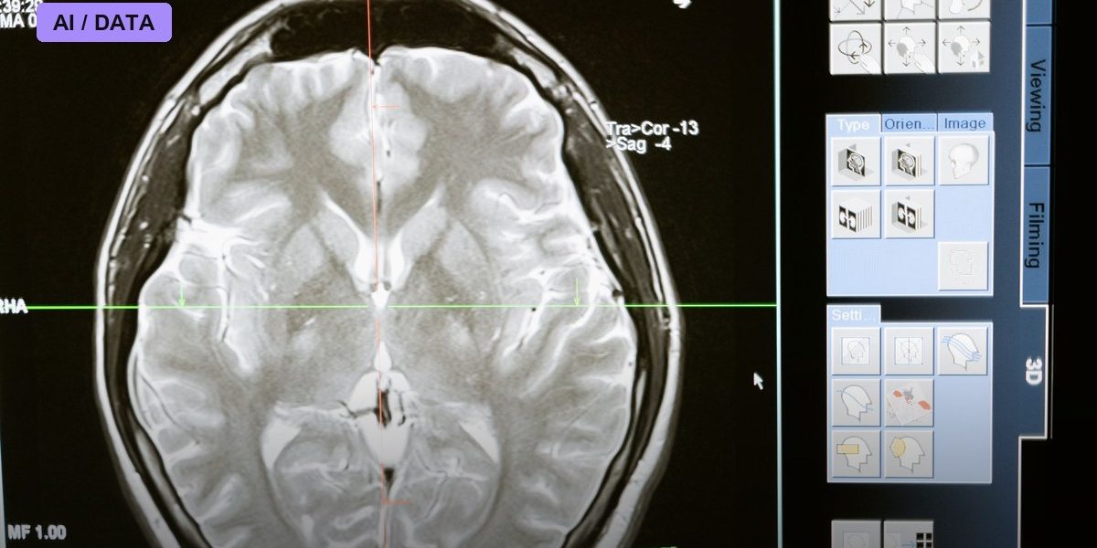
An AI system that scans ordinary, contrast-free brain CT images can spot dangerous stroke-causing blockages that clinicians alone frequently miss, according to a multinational validation study led by Dr. Chi Kyung Kim of the Department of Neurology at Korea University Guro Hospital. The research, carried out with colleagues including Dr. Kim Beom-joon of Korea University Guro Hospital’s neurology department and Dr. Jun Sun-woo of the radiology department at Seoul National University Bundang Hospital, tested an algorithm built by the Korean medical-imaging company JLK against real emergency-room cases from Korea and the United States, and the results were published in the Journal of NeuroInterventional Surgery.
The Problem the Study Set Out to Fix
Large vessel occlusion, or LVO, is among the most severe forms of ischemic stroke, caused when a major brain artery is blocked and a substantial portion of brain tissue is starved of blood flow. Patients with LVO generally need a mechanical thrombectomy, a procedure to physically remove the clot, and outcomes depend heavily on how fast it happens, a window clinicians call the golden hour. The gold-standard way to confirm an LVO is CT angiography or MRI, both of which require contrast dye, specialized protocols, and often a radiologist or neurologist with subspecialty training to interpret quickly, resources that are not evenly available at every hospital an ambulance might reach first. Plain, non-contrast CT is faster, cheaper, and available almost everywhere, but reading it for subtle signs of LVO is notoriously difficult even for experienced clinicians.
What the Data Showed
The researchers analyzed a multinational cohort of 963 patients, 723 from Korea and 240 from the United States. In a reader study, clinicians interpreting non-contrast CT scans without AI assistance achieved an area under the curve of 0.718, correctly flagging LVO with a sensitivity of just 46.6 percent, though specificity was high at 91.9 percent, meaning they rarely raised false alarms but missed more than half of true cases. When the same clinicians used the AI as a second reader, performance jumped to an area under the curve of 0.852, with sensitivity rising to 63.7 percent and specificity improving slightly to 94.9 percent. In practical terms, the research team calculated that for every 18 non-contrast CT scans read by medical staff, AI assistance identifies one additional LVO patient who would otherwise have been missed. In a separate validation of the algorithm’s standalone diagnostic performance across the two countries, the AI achieved an area under the curve of 0.963 in the Korean cohort and 0.899 in the U.S. cohort, indicating strong but not identical performance across health systems and scanner populations.
Guarding Against a New Kind of Error
One finding the researchers highlighted specifically was the absence of automation bias, the well-documented risk that clinicians start deferring to an algorithm’s judgment even when their own read would have been more accurate. The study reported that participating clinicians retained their own critical judgment while treating the AI’s output as a safety net rather than a final answer, a distinction that matters because automation bias has undermined the real-world value of decision-support tools in other areas of medicine even when the underlying algorithm tested well in the lab.
A Company Betting on Faster, Cheaper Imaging
JLK, the company behind the algorithm, has been pushing to get non-contrast CT-based stroke detection into wider clinical use, and has separately obtained U.S. Food and Drug Administration 510(k) clearance for an NCCT-based AI stroke detection tool, a regulatory step that allows the product to be marketed for clinical use in the United States. The company’s argument is that because plain CT scanners are already standard equipment in nearly every emergency department, an AI layer that upgrades their diagnostic value could extend LVO detection capability to smaller, rural, or resource-limited hospitals that lack a stroke neurologist or interventional radiologist on call around the clock.
The Limits Critics Point To
Even with AI assistance, a sensitivity of 63.7 percent means the tool still misses more than a third of true LVO cases, underscoring that researchers frame it as a triage aid rather than a replacement for definitive contrast imaging once a patient reaches a comprehensive stroke center. AI diagnostic researchers more broadly have also cautioned that algorithms validated on specific scanner models and patient populations, as this one was across Korean and American cohorts, can perform unpredictably when deployed on equipment or demographics not represented in the training data, which is part of why the gap between the Korean AUC of 0.963 and the U.S. AUC of 0.899 is being watched closely rather than dismissed as noise.
What’s Next
With FDA clearance already secured for the non-contrast approach, the near-term question is how quickly hospital systems, particularly smaller and rural ones without dedicated neuroradiology coverage, choose to adopt it, and whether real-world deployment preserves the sensitivity gains seen in this controlled study. Stroke researchers say the next round of validation will need to track actual treatment-time and outcome data, not just diagnostic accuracy, to prove that catching more LVO cases on a first scan translates into more patients reaching a thrombectomy suite inside the golden hour.
Photo: MART PRODUCTION / PEXELS via Pexels