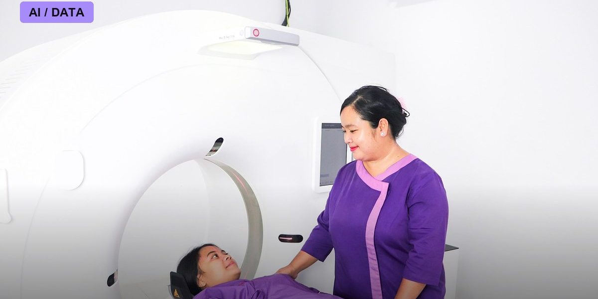
A research team led by Dr. Chi Kyung Kim of Korea University Guro Hospital has completed a multinational validation of an AI system that can detect large vessel occlusion, the type of blood clot responsible for the most disabling strokes, using nothing more than a standard, non-contrast brain CT scan. The findings, published in the Journal of NeuroInterventional Surgery and reported by Medical Xpress in September 2026, address a practical bottleneck in stroke care: the imaging that most reliably shows a major clot usually requires injecting contrast dye and running specialized software, resources that are not available around the clock at every hospital that sees stroke patients.
Why non-contrast CT is the harder, more useful target
When someone arrives at an emergency room with stroke symptoms, the fastest imaging option is almost always a plain CT scan without contrast dye, because it does not require the extra time, equipment, or the trained staff needed to administer and time a contrast injection. The problem is that a plain CT scan makes it much harder to see whether a large vessel is actually blocked; that determination has traditionally relied on CT angiography or CT perfusion imaging, both of which use contrast dye and are not always immediately available, especially at smaller or rural hospitals overnight. Dr. Kim’s team trained and tested their AI model specifically to work from the plain, dye-free scan that virtually every hospital can already perform, rather than requiring the more specialized imaging that many cannot.
How the validation was structured
The study describes itself as a multinational validation, meaning the AI model was tested against non-contrast CT scans and confirmed large vessel occlusion diagnoses drawn from multiple countries and hospital systems, rather than a single institution’s patient population. That kind of cross-border testing matters in stroke AI specifically because CT scanner hardware, patient demographics, and even how strokes present clinically can vary meaningfully between health systems, and models trained narrowly on one hospital’s data have previously struggled when applied elsewhere. According to the research team’s reporting, the model performed accurately at precisely detecting large vessel occlusion across these varied settings using only the widely available scan type.
The stakes of getting stroke triage right
Large vessel occlusion strokes are typically treated with mechanical thrombectomy, a procedure in which a specialist threads a device through the patient’s blood vessels to physically remove the clot, but that procedure can usually only be performed at specialized stroke centers. Determining quickly whether a patient has this type of clot decides whether they need to be transferred urgently to such a center, and every delay in that decision costs brain tissue: physicians commonly cite estimates of roughly 1.9 million neurons lost per minute in an ongoing large vessel occlusion. A tool that can flag this specific type of stroke from a scan every hospital already has the equipment to run could, in principle, speed up transfer decisions at hospitals that currently have to either guess, wait for a specialist read, or default to sending every possible case for advanced imaging that isn’t readily available.
What clinicians outside the study are watching for
Stroke and health-IT researchers, including those cited in a companion AuntMinnie analysis of hospital resourcing and AI stroke tools published in 2026, caution that detecting a clot on a scan is only the first link in a longer chain that determines patient outcomes. Even a highly accurate AI flag is only useful if the receiving hospital has a thrombectomy-capable team available to act on it, ambulance or helicopter transfer capacity to move the patient quickly, and staff trained to trust and act on the AI’s output rather than waiting for a second confirmatory read. Researchers in the field have noted that hospitals with weaker existing stroke protocols tend to see smaller real-world gains from adding AI, even when the underlying detection accuracy is strong, because the bottleneck shifts elsewhere in the care pathway.
What’s next
Dr. Kim’s team says the multinational results are intended to support revisions to international guidelines on which AI stroke-imaging tools can be relied upon without contrast imaging, a change that could make it easier for smaller and rural hospitals, which often lack round-the-clock contrast-imaging capability, to adopt fast AI-based stroke triage. If those guideline changes follow, health systems such as OSF HealthCare, which has separately been expanding AI stroke-imaging software across its own hospital network in Illinois and Michigan this September, could have a scientific basis for extending similar tools to sites that currently rely on plain CT scans alone.
Photo: goesstock / PIXABAY via Pixabay